Echocardiogram vs. EKG/ECG: What’s the Difference?

Introduction
Echocardiograms and EKG’s are two of the most common tests to diagnose various heart conditions. While there are similarities between these non-invasive tests, there are key differences to consider when a patient is experiencing symptoms. In this article, we’ll explain the difference between an EKG and an echocardiogram.
What is an EKG/ECG?
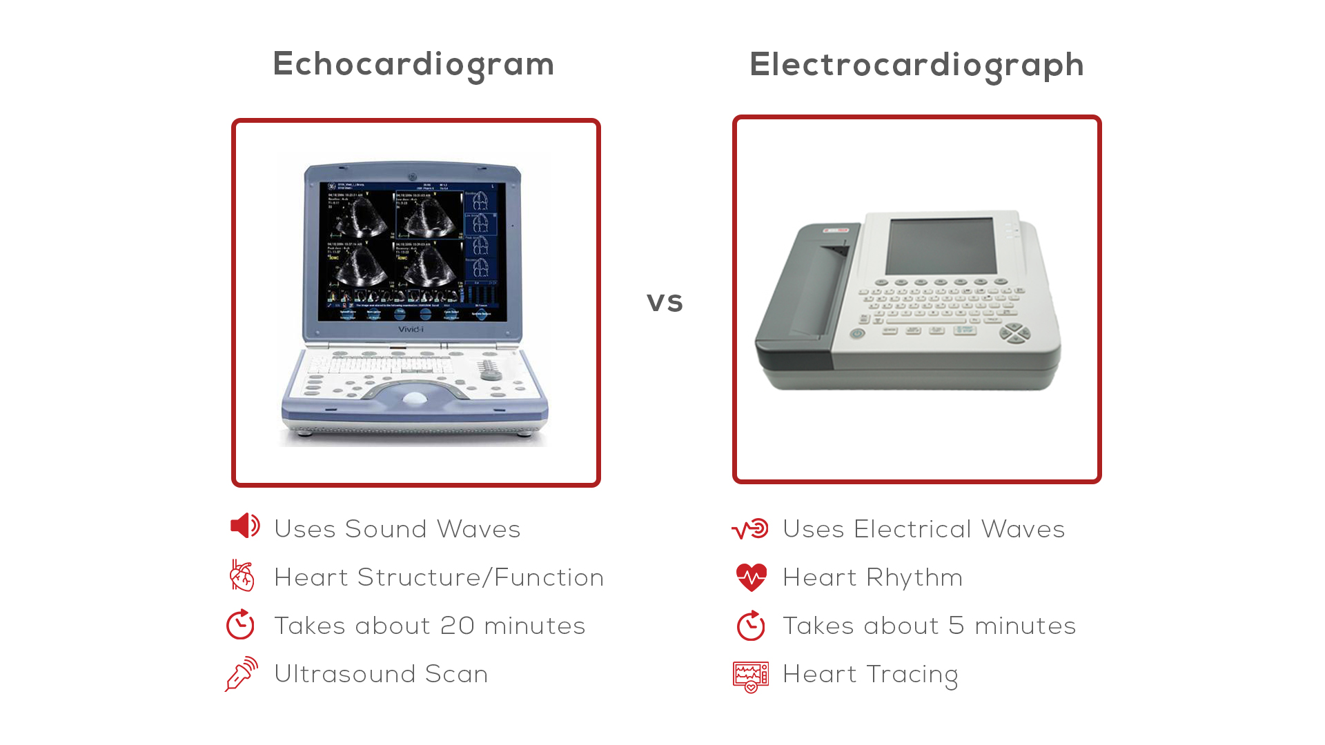
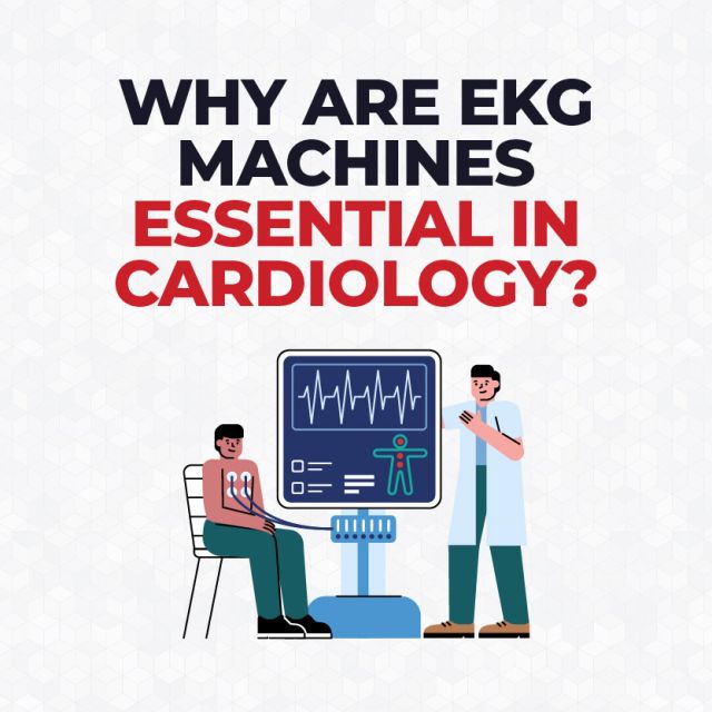
An EKG/ECG, or electrocardiogram, is a non-invasive test used to detect and monitor heart conditions and diseases. EKG/ECG tests are performed using an EKG/ECG machines to record the electrical signals in the heart. The electrodes attach to the patient’s body in specific positions to ensure accurate reading. An EKG test takes just a few minutes.
What is an Echocardiogram?
An echocardiogram provides an ultrasound image of the heart, including its valves and chambers. Echocardiograms show the heart chamber sizes and pumping function to identify heart disease and diagnose heart failure. 2D/3D ultrasounds are typically used to create these photos. An echocardiogram can take anywhere from 15 to 60 minutes.
What is the Difference Between an EKG and an Echocardiogram?
An electrocardiogram (ECG or EKG) and an echocardiogram (echo) are both commonly used tests to evaluate heart health, but they measure very different things and provide different visuals.
An ECG records the electrical activity of the heart using electrodes placed on the skin. EKG tests provide information about heart rhythm, heart rate, and can help detect arrhythmias and heart attacks.
In contrast, an echocardiogram is an ultrasound-based imaging test that shows detailed pictures of the heart’s structure and function. Echocardiograms assess the heart’s chambers, valves, pumping strength, and blood flow to diagnosing conditions such heart failure.


